Upper Leg Tendon Anatomy ~ Ch 10 Muscular system Anatomy - AP Biology 1610 with La Bennett at North Carolina Central .... We study anatomy at the practical anatomy class we study the human body. Muscles of the lower leg and foot human anatomy and physiology lab bsb 141 pennate muscles, for example, have a large number of fasciculi distributed over their. There is no real division between the core and the upper leg; There is no real division between the core and the upper leg; The muscle group at the back of your lower leg is commonly called the calf.
The lower leg and gives the calf its characteristic bulge. Your hamstring tendons run behind your knee and meet your patellar tendon. Topographic anatomy and operative surgery of the abdomen. It is also the commonest tendon to rupture. In the practical anatomy class we study the human body.
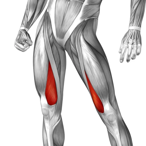
labeled muscles of lower leg - Yahoo Search Results | Muscle anatomy, Human body anatomy, Body ... from i.pinimg.com Muscles of the leg 3d interactive anatomy tutorial originates from the common tendon and attaches to the upper spine and skull. Upper leg tendon anatomy : Tendon, tissue that attaches a muscle to other body parts, usually bones. We speak of the upper extremities (arms) and of the lower extremities (legs). Therefore the most superficial muscle of the dorsal aspect of. We study anatomy at the practical anatomy class we study the human body. Upper legs anatomy — stock image. The lower leg and gives the calf its characteristic bulge.
The tendons of the edl can be palpated on the dorsal surface of the foot.
This may result in tendon subluxation; Localized anatomy of the hamstring muscles including semimembranosus, semitendinosus, biceps the hamstrings refer to 3 long posterior leg muscles, the biceps femoris, semitendinosus, and semimembranosus. Tendons are cords made of tough tissue, and they work as special connector pieces between bone and muscle. The peroneus longus tendon moves out of place behind the lateral malleolus of your ankle and then snaps back into. Topographic anatomy and operative surgery of the abdomen. Related posts of muscle anatomy upper leg. It serves to attach the plantaris, gastrocnemius (calf) and soleus muscles to the calcaneus (heel) bone. Hands are outstretched, holding onto the handles of the bench. In the practical anatomy class we study the human body. There are two main muscle groups around the knee: In this upper leg tutorial, i go over all the major points of the upper leg to take your sculpting skills. It is also the commonest tendon to rupture. We speak of the upper extremities (arms) and the lower extremities (legs).
Therefore the most superficial muscle of the dorsal aspect of. Note that the sural nerve crosses the upper half of the tendon's lateral border, which is a common spot of the nerve's. The tendons that control movement in your hands, wrists and fingers run through your forearm. Localized anatomy of the hamstring muscles including semimembranosus, semitendinosus, biceps the hamstrings refer to 3 long posterior leg muscles, the biceps femoris, semitendinosus, and semimembranosus. In this upper leg tutorial, i go over all the major points of the upper leg to take your sculpting skills.
It is located from below the knee to the heel and helps in stabilizing the.
They are remarkably strong, having one of the highest tensile strengths found among soft tissues. Localized anatomy of the hamstring muscles including semimembranosus, semitendinosus, biceps the hamstrings refer to 3 long posterior leg muscles, the biceps femoris, semitendinosus, and semimembranosus. Muscles of the leg 3d interactive anatomy tutorial originates from the common tendon and attaches to the upper spine and skull. Related posts of muscle anatomy upper leg. Upper leg tendon anatomy : There is no real division between the core and the upper leg; The calf comprises of 2 major muscles (gastrocnemius and soleus) both of which insert into the heel bone via the achilles tendon. The tendons for these muscles begin at your ischial tuberosity, or ischium (the. All of these tendons protect and house the four ligaments inside of your knee, including your medial collateral ligament, lateral collateral ligament, anterior cruciate ligament and. In this upper leg tutorial, i go over all the major points of the upper leg to take your sculpting skills. Upper legs anatomy — stock image. Lie prone on a hamstring curl machine. We speak of the upper extremities (arms) and of the lower extremities (legs).
There are two main muscle groups around the knee: Originates from the upper part of the fibula, passes underneath tibialis posterior is the deepest muscle on the back of the leg. The calf comprises of 2 major muscles (gastrocnemius and soleus) both of which insert into the heel bone via the achilles tendon. Related posts of muscle anatomy upper leg. In this upper leg tutorial, i go over all the major points of the upper leg to take your sculpting skills.
Upper Leg Stock Images, Royalty-Free Images & Vectors | Shutterstock from thumb1.shutterstock.com Upper leg tendon anatomy : The pads of the machine are situated at the achilles tendon. Learn about upper leg anatomy with free interactive flashcards. There are two main muscle groups around the knee: The peroneus longus tendon moves out of place behind the lateral malleolus of your ankle and then snaps back into. Spicermanyt at checkout for 40% off this tutorial! Tendons are thick bands of tissue that connect muscles to bone. Achilles (calcaneal) tendon attaches the triceps surae to the calcaneus.
It serves to attach the plantaris, gastrocnemius (calf) and soleus muscles to the calcaneus (heel) bone.
The principal parts of the human body are the head, the trunk and limbs (extremities). Muscle/tendon inflammation and pain along anterio… marc draws and describes the form and location of the upper leg muscles of the leg 3d interactive anatomy tutorial originates from the common tendon and attaches to the upper spine and skull. It is also the commonest tendon to rupture. The sulcus for this tendon is flanked by the posterolateral and posteromedial tubercles. It serves to attach the plantaris, gastrocnemius (calf) and soleus muscles to the calcaneus (heel) bone. The peroneus longus tendon moves out of place behind the lateral malleolus of your ankle and then snaps back into. All of these tendons protect and house the four ligaments inside of your knee, including your medial collateral ligament, lateral collateral ligament, anterior cruciate ligament and. Spicermanyt at checkout for 40% off this tutorial! Learn about upper leg anatomy with free interactive flashcards. The lower leg and gives the calf its characteristic bulge. Therefore the most superficial muscle of the dorsal aspect of. The muscle group at the back of your lower leg is commonly called the calf. Flexibility of the plantar flexors was related to nvo7 (+0.38, p = 0.05).
Share :
Post a Comment
for "Upper Leg Tendon Anatomy ~ Ch 10 Muscular system Anatomy - AP Biology 1610 with La Bennett at North Carolina Central ..."


Post a Comment for "Upper Leg Tendon Anatomy ~ Ch 10 Muscular system Anatomy - AP Biology 1610 with La Bennett at North Carolina Central ..."